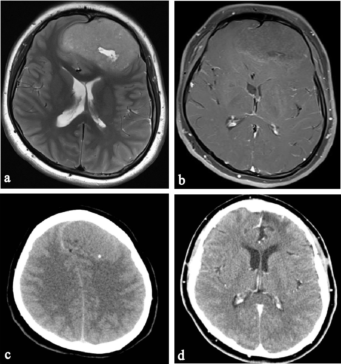
Figure 1. Preoperative MRI, CT and postoperative CT. (a) Cystic component at a tumor center of frontal convexity. (b) Heterogeneous enhancement of tumor after IV gadolinium. (c) Spot-like calcification of posterior tumor margin. (d) Tumor recurrence is not found on enhanced axial CT image 4 years post surgery.
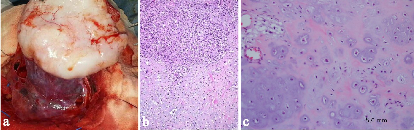
Figure 2. Intraoperative photograph and histological findings. (a) The tumor margin is clear without invasion. (b) The specimen showing a biomorphic appearance-area of well-differentiated hyaline cartilage juxtaposed to undifferentiated stroma (H&E × 100). (c) The cartilaginous components are composed of atypical chondrocytes with nuclear pleomorphism and mitosis in lacunae (H&E × 200).