Figures
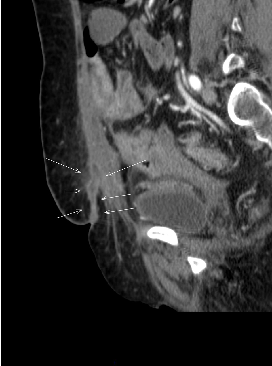
Figure 1. Abdominopelvic CT scan of sagittal view with IV and PO contrast (white arrows) showing fistulous tract and enterocutaneous fistula.
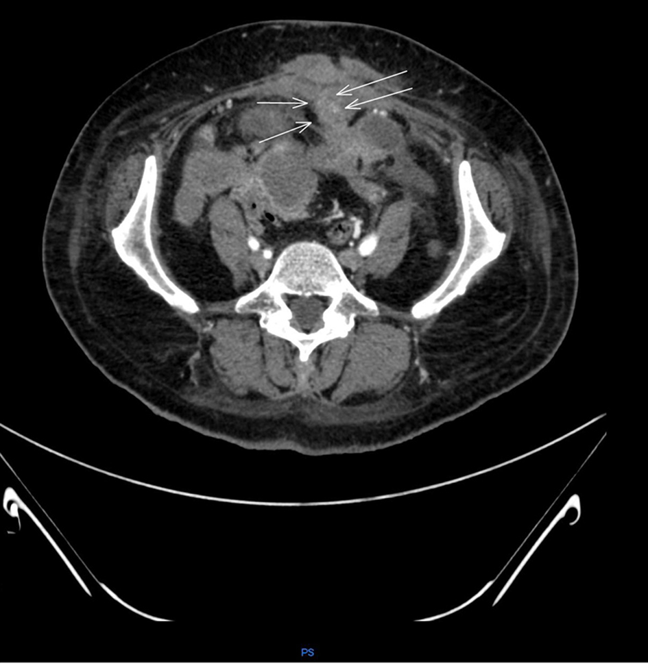
Figure 2. Abdominopelvic CT scan with IV and PO contrast transverse view (white arrows) pointing fistulous tract and enterocutaneous fistula.
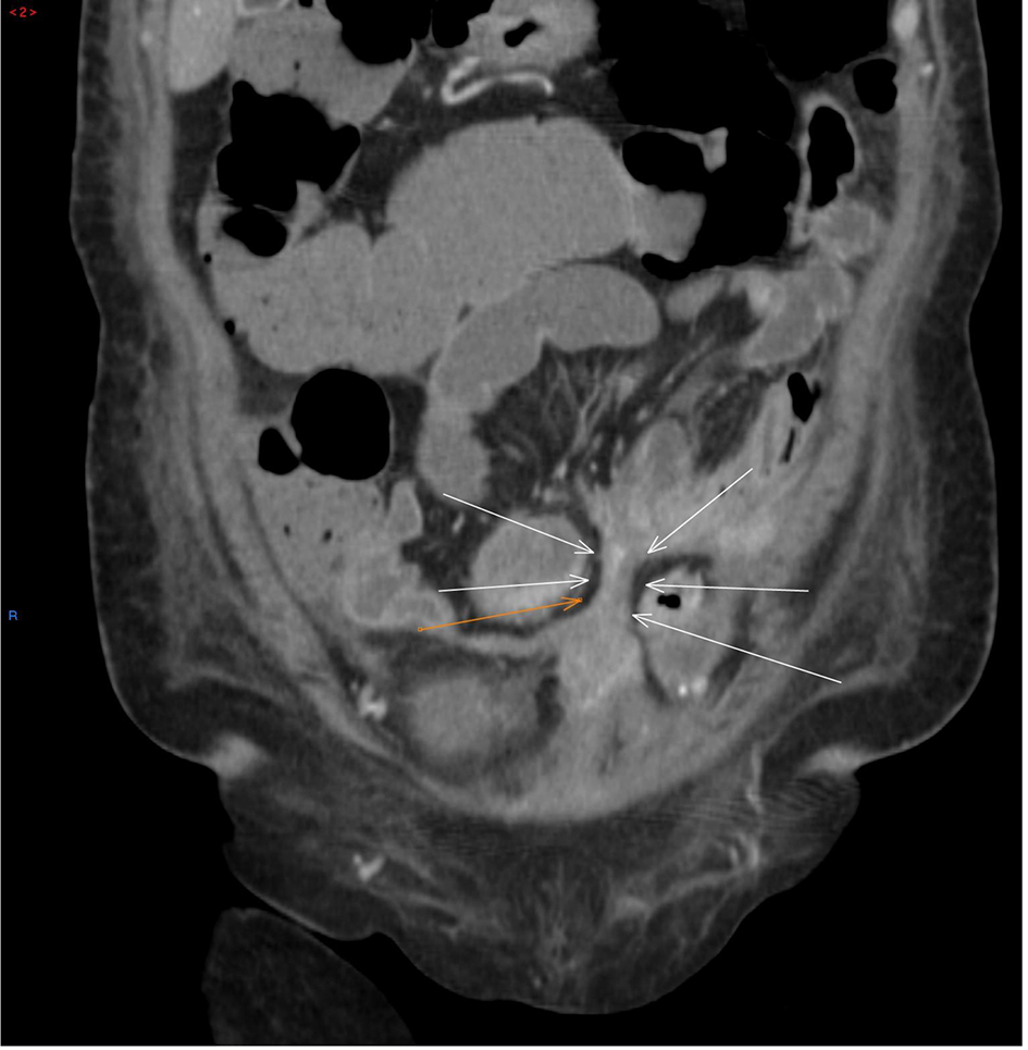
Figure 3. Abdominopelvic CT scan with PO and IV contrast coronal view showing colonic fistulous tract (white and orange arrows).
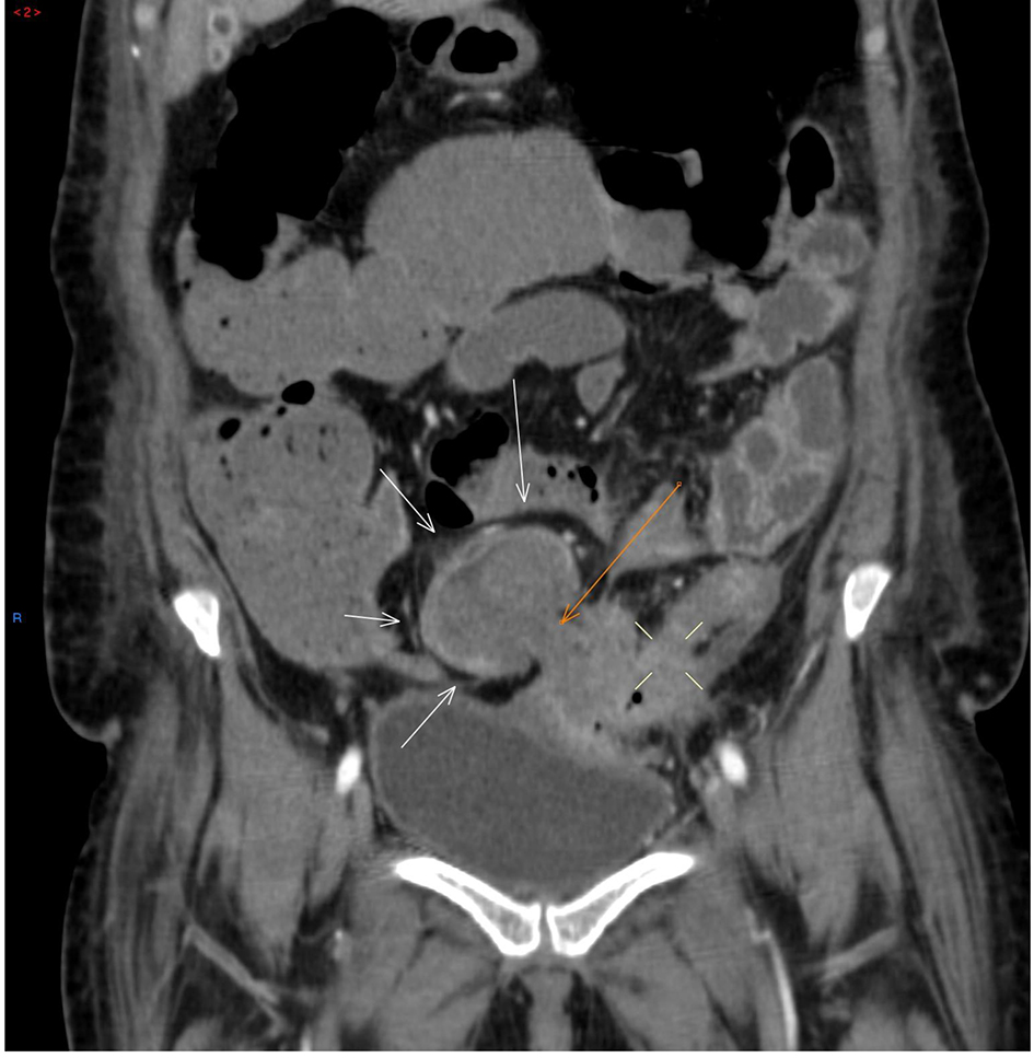
Figure 4. Abdominopelvic CT scan with PO and IV contrast coronal view showing large intraabdominal abscess (white arrows, orange arrow) showing bowel wall (four white lines showing another intraabdominal abscess).
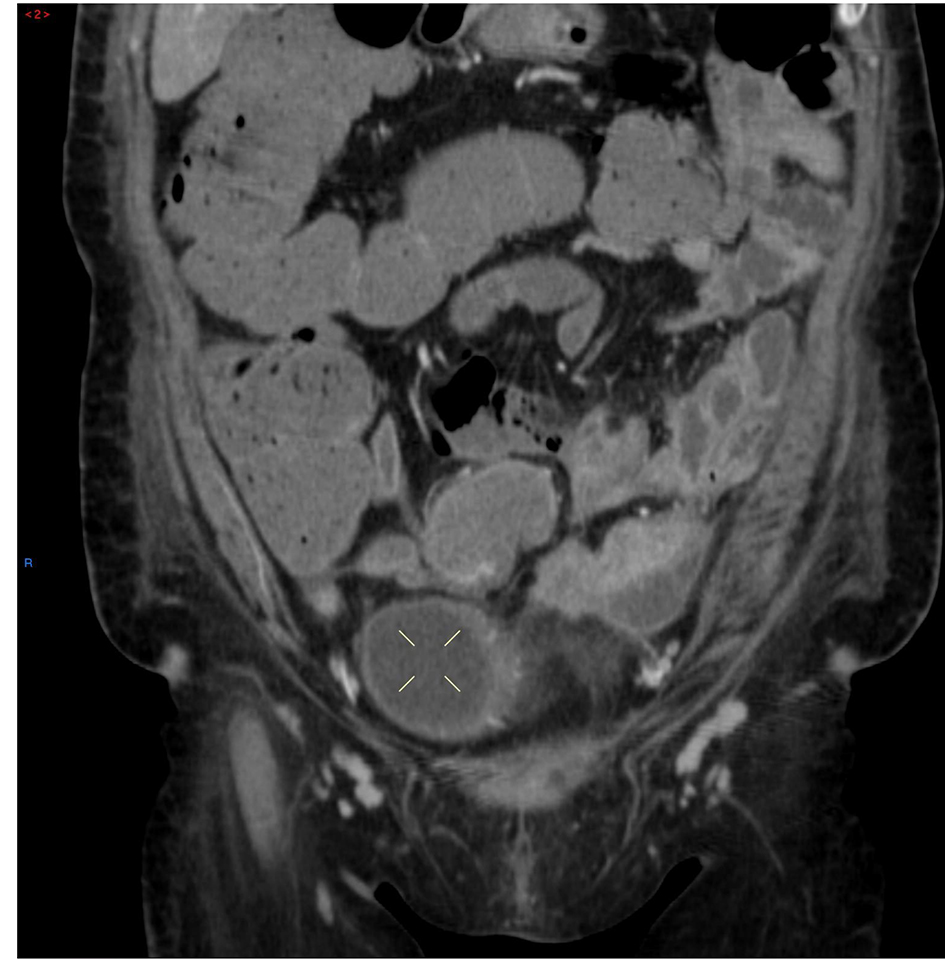
Figure 5. Coronal abdominopelvic CT scan with IV and PO contrast (four white lines) showing large intraabdominal abscess.
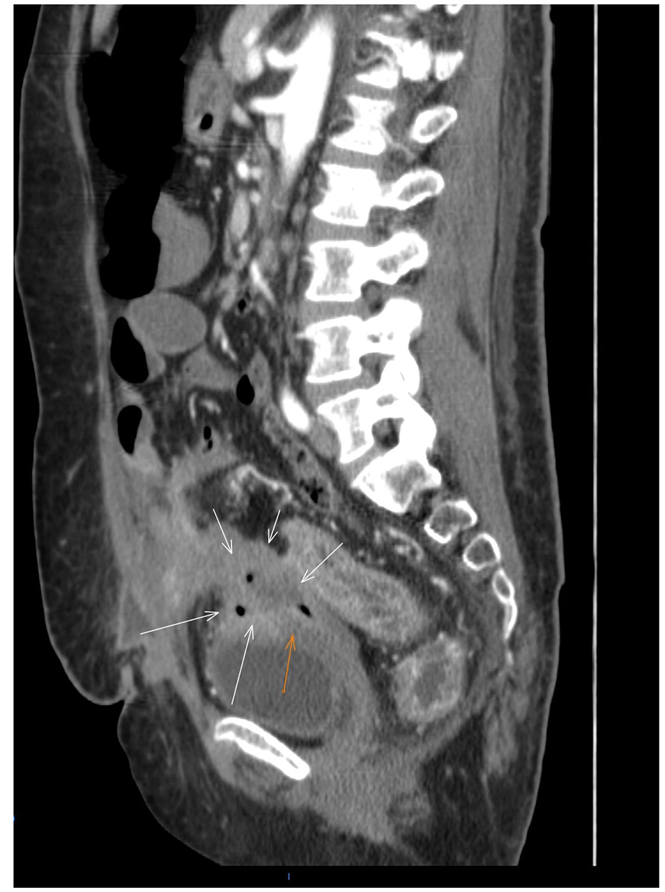
Figure 6. Abdominopelvic CT scan with IV and PO contrast sagittal view (white arrows) showing fistulizing tract and (orange arrow) showing rectovaginal fistula.
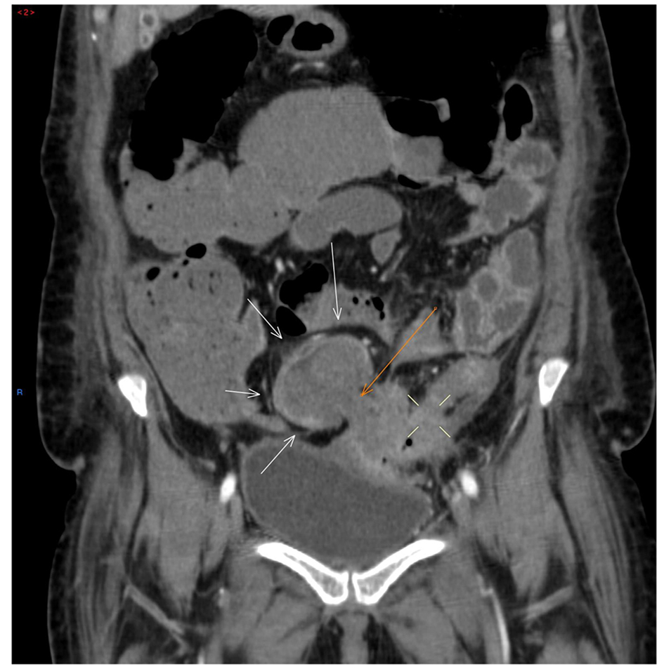
Figure 7. Abdominopelvic CT scan with PO and IV contrast coronal view showing colonic fistulous tract (white and orange arrows).