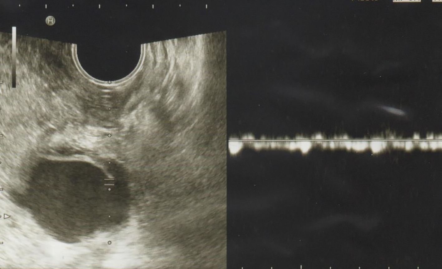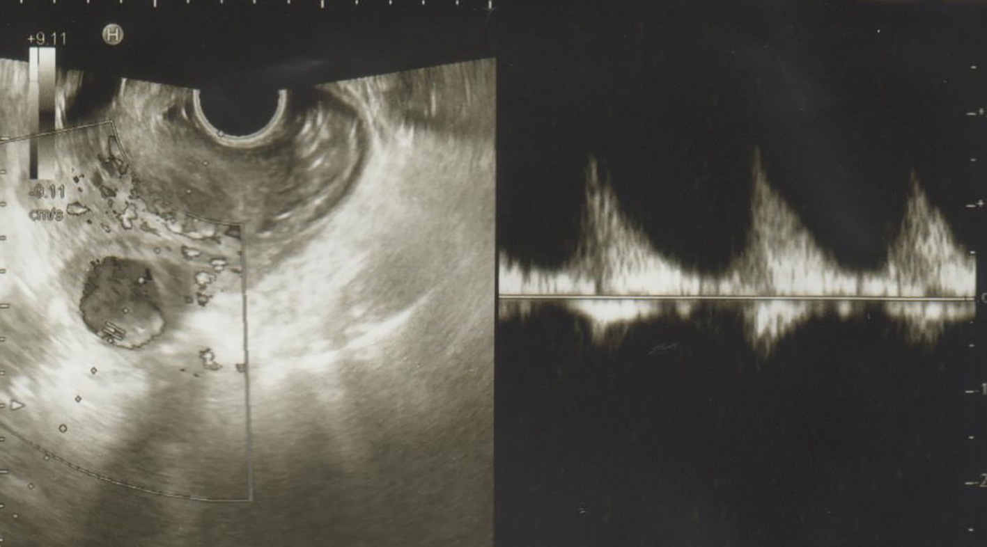
Figure 1. Transvaginal ultrasonography showing the presence of low density cyst with a diameter of 4 cm with spiral pulsating blood flow in the cavity of the cyst in the vicinity of the ascending branches of the left uterine artery.
| Journal of Medical Cases, ISSN 1923-4155 print, 1923-4163 online, Open Access |
| Article copyright, the authors; Journal compilation copyright, J Med Cases and Elmer Press Inc |
| Journal website http://www.journalmc.org |
Case Report
Volume 8, Number 8, August 2017, pages 241-242
Ultrasonographic Findings of Uterine Artery Pseudoaneurysm
Figures