Journal of Medical Cases, ISSN 1923-4155 print, 1923-4163 online, Open Access
Article copyright, the authors; Journal compilation copyright, J Med Cases and Elmer Press Inc
Journal website http://www.journalmc.org

Case Report

Volume 7, Number 12, December 2016, pages 531-535


Cardiac Amyloidosis Presenting With Myocardial Ischemia: Case Report and Review Article

Figures

Figure 1.
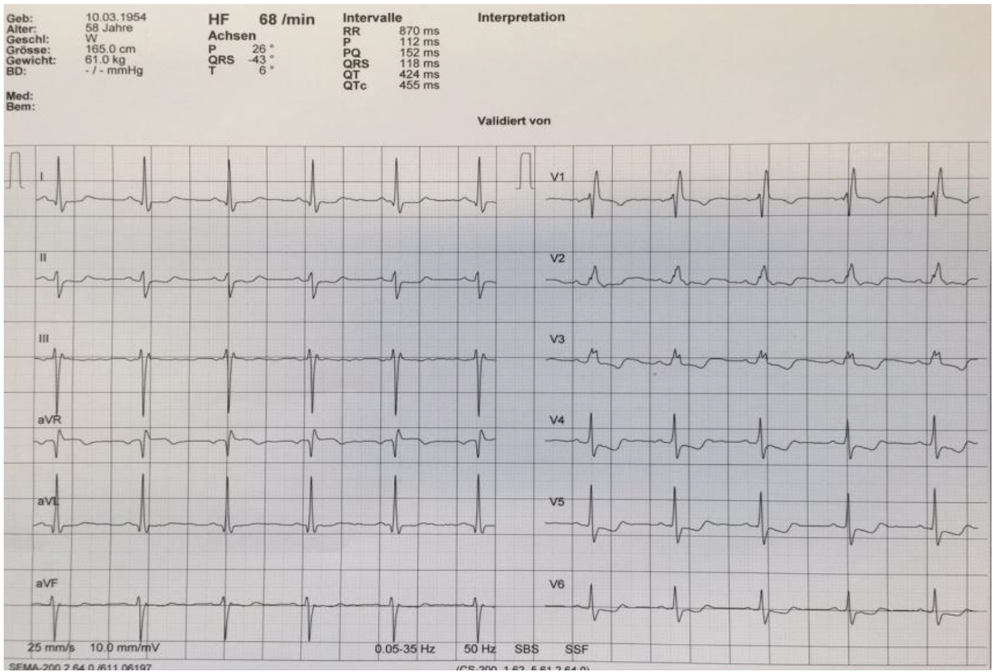
Figure 1. Electrocardiogram reveals a right bundle branch block and left anterior fascicular block, in addition T-wave inversion in leads V3-V5.
Figure 2.
Figure 2. Coronary angiography showing normal left and right coronary arteries.
Figure 3.
Figure 3. MRI adenosine stress test showing diffuse endocardial perfusion delay.
Figure 4.
Figure 4. Endomyocardial biopsy: the amyloid deposits turn red in the Congo red staining (left); same cut of Congo red staining in polarized light; abnormal green to yellow birefringence of amyloid deposits (right).
Figure 5.
Figure 5. Postmortem endomyocardial histological samples show abundant interstitial, subendocardial (left) and small artery media (right) deposits of amyloid substance.