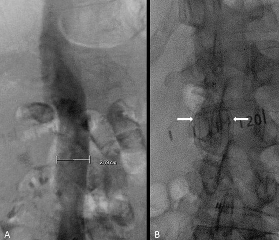
Figure 1. (A) Inferior vena cavogram showing a diameter of approximately 21 mm. (B) Deployed TrapEase filter (between arrows).
| Journal of Medical Cases, ISSN 1923-4155 print, 1923-4163 online, Open Access |
| Article copyright, the authors; Journal compilation copyright, J Med Cases and Elmer Press Inc |
| Journal website http://www.journalmc.org |
Case Report
Volume 2, Number 5, October 2011, pages 201-205
Inferior Vena Cava Filter Migration to the Right Ventricle: A Case Report and Review of Filter Migration and Misdeployment
Figures