Figures

Figure 1. Metastatic lesions (arrows) in the left ilium, ischium, and femur.

Figure 2. Intraoral photograph showing exposed bone (arrow) in the lingual posterior area of the right mandible.

Figure 3. Panoramic radiograph at revisit showing a slightly widened periodontal ligament space in the tooth cervix, periodontal alveolar bone resorption in the area surrounding the right lower second molar, and sclerosis of cancellous bone in the molar region of the right mandible.
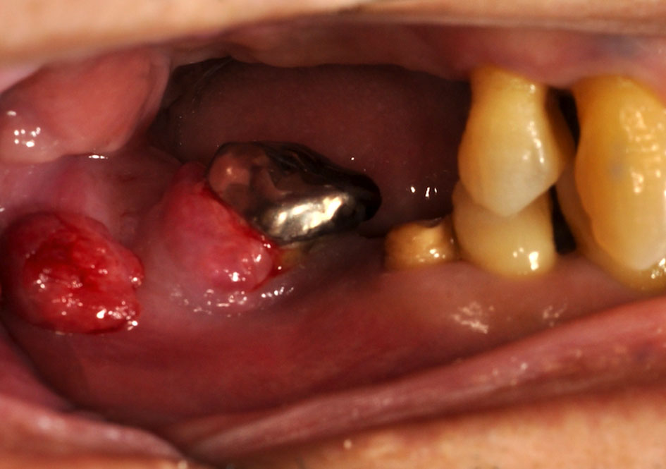
Figure 4. Intraoral photograph showing a fistula, with gingival redness and swelling, on the buccal gingiva of the mandibular right second molar.

Figure 5. CT images immediately before sequestrectomy reveal characteristic findings of chronic mandibular osteomyelitis.

Figure 6. MR images of the right mandibular lesion and bone metastasis in the left femur. (a) Metastatic tumor in the left femur (arrow). (b) Pathological change in the bone marrow of the right mandible (arrow).

Figure 7. Histopathological specimen obtained during sequestrectomy (hematoxylin and eosin, × 4).