Journal of Medical Cases, ISSN 1923-4155 print, 1923-4163 online, Open Access
Article copyright, the authors; Journal compilation copyright, J Med Cases and Elmer Press Inc
Journal website http://www.journalmc.org

Case Report

Volume 6, Number 5, May 2015, pages 211-213


Orbital Cavernous Hemangioma With Proptosis in a Child

Figures

Figure 1.
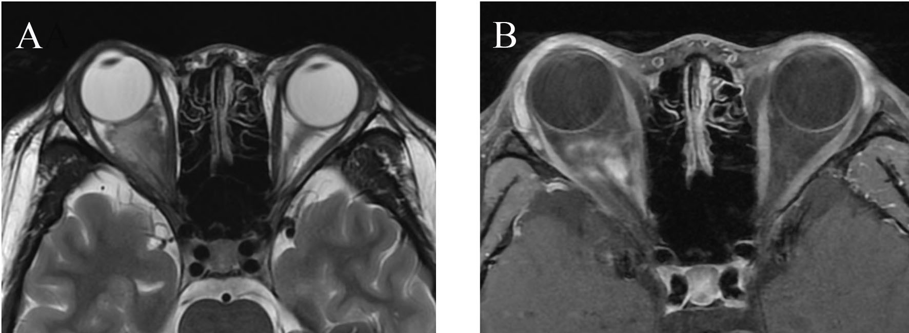
Figure 1. Axial T2-weighted image (A) and postcontrast T1-weighted image (B). A slightly irregular large intraconal mass of pyramidal shape leads to proptosis. The extraocular muscles are displaced and stretched. The irregular enhancement inside the cavernous hemangioma is seen.
Figure 2.
Figure 2. Histological examination revealing a cavernous hemangioma (H&E staining, original magnification × 200).
Figure 3.
Figure 3. A CT showing a large soft tissue mass in the mediosuperior compartment of right orbit. The cavernous hemangioma encases the right optic nerve.