
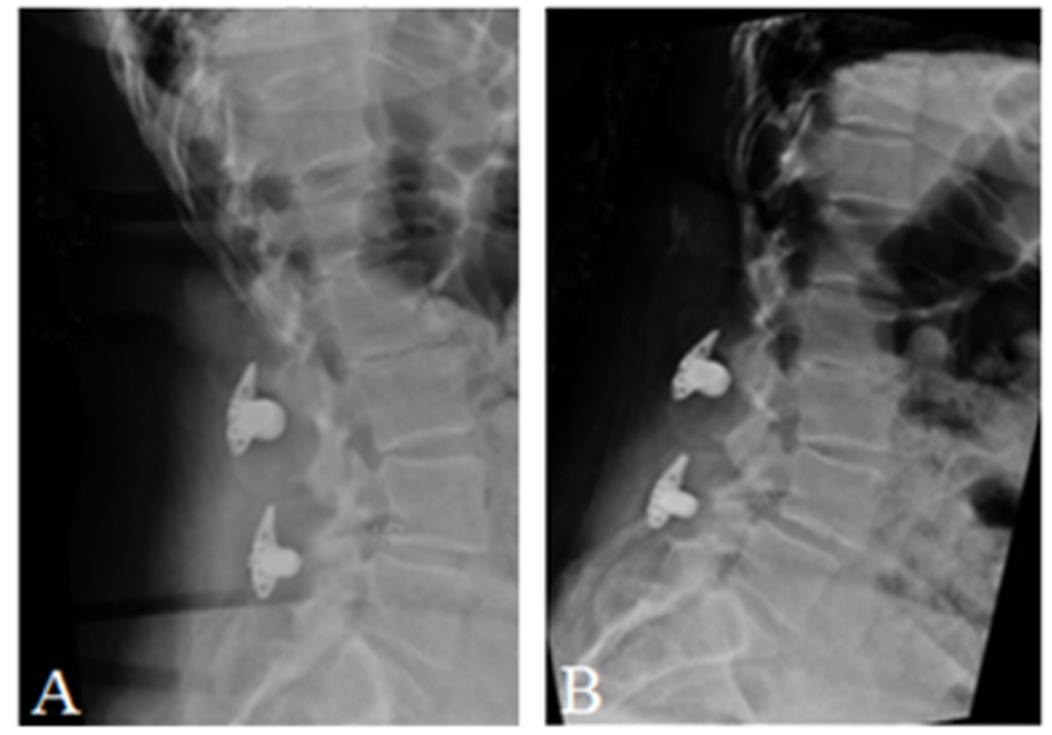
Figure 1. Schematic of X-STOP device.
| Journal of Medical Cases, ISSN 1923-4155 print, 1923-4163 online, Open Access |
| Article copyright, the authors; Journal compilation copyright, J Med Cases and Elmer Press Inc |
| Journal website http://www.journalmc.org |
Case Report
Volume 6, Number 5, May 2015, pages 205-210
A New Approach to Quantify Functional Improvements Following X-Stop Spacer Procedure: A Case Report
Figures


Table
| PROMIS-29 domain | Pre-op t-score (SE) | Post-op t-score (SE) | Change |
|---|---|---|---|
| Physical function | 43.4 (2.4) | 56.9 (6.7) | Significantly improved |
| Anxiety | 51.2 (3.1) | 51.2 (3.1) | Unchanged |
| Depression | 41.0 (6.2) | 41.0 (6.2) | Unchanged |
| Fatigue | 60.7 (2.3) | 48.6 (2.5) | Significantly improved |
| Sleep disturbance | 32.0 (5.2) | 32.0 (5.2) | Unchanged |
| Satisfaction with social role | 38.8 (2.1) | 64.1 (5.1) | Significantly improved |
| Pain interference | 63.8 (1.8) | 55.6 (1.9) | Significantly improved |
| Pain intensity | 7/10 | 6/10 | Improved |