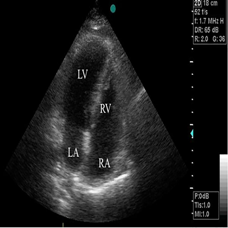
Figure 1. Apical four-chamber view showing the right atrial thrombus (arrow) and the enlargement of the right heart chambers. RA: right atrium; RV: right ventricle.
| Journal of Medical Cases, ISSN 1923-4155 print, 1923-4163 online, Open Access |
| Article copyright, the authors; Journal compilation copyright, J Med Cases and Elmer Press Inc |
| Journal website http://www.journalmc.org |
Case Report
Volume 5, Number 10, October 2014, pages 532-534
Massive Pulmonary Embolism as a Complication of Coronary Angiography and Its Successful Treatment With Half-Dose Alteplase
Figures