Journal of Medical Cases, ISSN 1923-4155 print, 1923-4163 online, Open Access
Article copyright, the authors; Journal compilation copyright, J Med Cases and Elmer Press Inc
Journal website http://www.journalmc.org

Case Report

Volume 5, Number 10, October 2014, pages 541-544


Colonoscopy-Induced Splenic Injury: An Unusual Complication Warranting Urgent Attention

Figure

Figure 1.
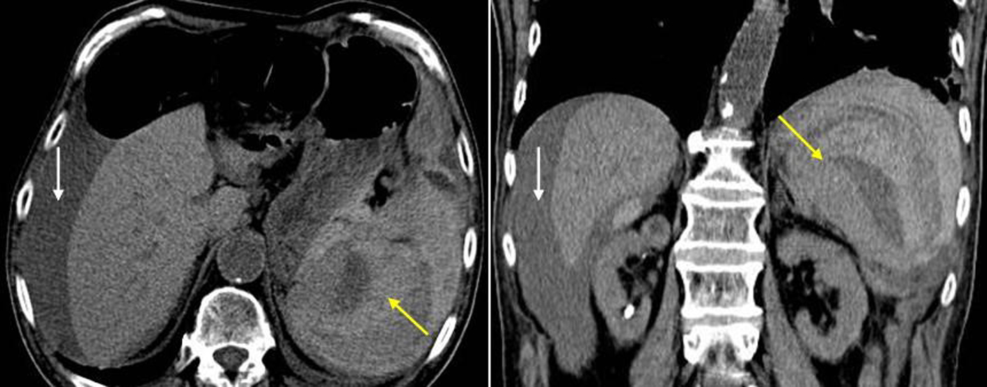
Figure 1. Non-contrast axial and coronal CT image of the abdomen shows a shattered spleen with a large amount of surrounding hemorrhage (yellow arrows) consistent with a grade 4-5 splenic injury. Hemoperitoneum extends to the perihepatic region (white arrows).

Table

Table 1. Risk Factors of Colonoscopy-Induced Splenic Injury [4, 5, 7, 15, 17-24]
 
Patient-dependent risk factorsEndoscopist-dependent risk factors
AdhesionsExcessive traction on the splenocolic ligament
Prior intra-abdominal surgeriesDirect injury to spleen
Inflammatory intra-abdominal processesCertain endoscopic maneuvers such as
Inflammatory bowel disease  Hooking the splenic flexure
Pancreatitis  Slide by advancement
Diverticulitis  Alpha maneuver
Splenomegaly  Straightening the sigmoid loop
Infectious disease  Intubation of ileum
Anticoagulation  Excessive looping
Neoplasm  External pressure
Multiple colonoscopies  Polypectomy
  Biopsy
  Technically difficult colonoscopy
  Supine position
  Operator inexperience