Journal of Medical Cases, ISSN 1923-4155 print, 1923-4163 online, Open Access
Article copyright, the authors; Journal compilation copyright, J Med Cases and Elmer Press Inc
Journal website http://www.journalmc.org

Case Report

Volume 5, Number 2, February 2014, pages 116-119


Sarcomatoid Carcinoma of the Urinary Bladder: A Case Report and Review of the Literature

Figures

Figure 1.
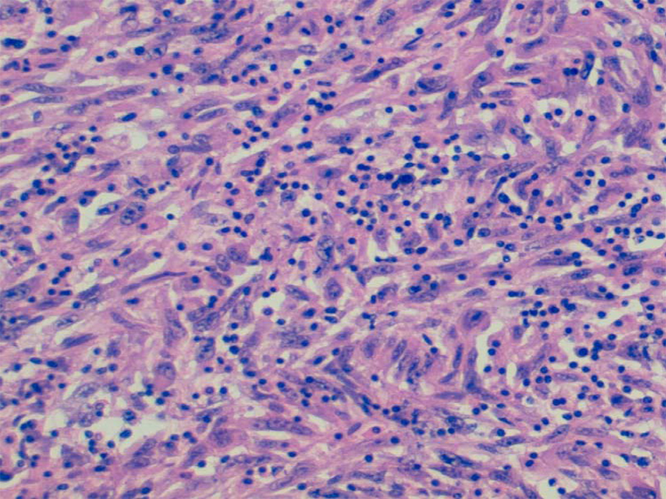
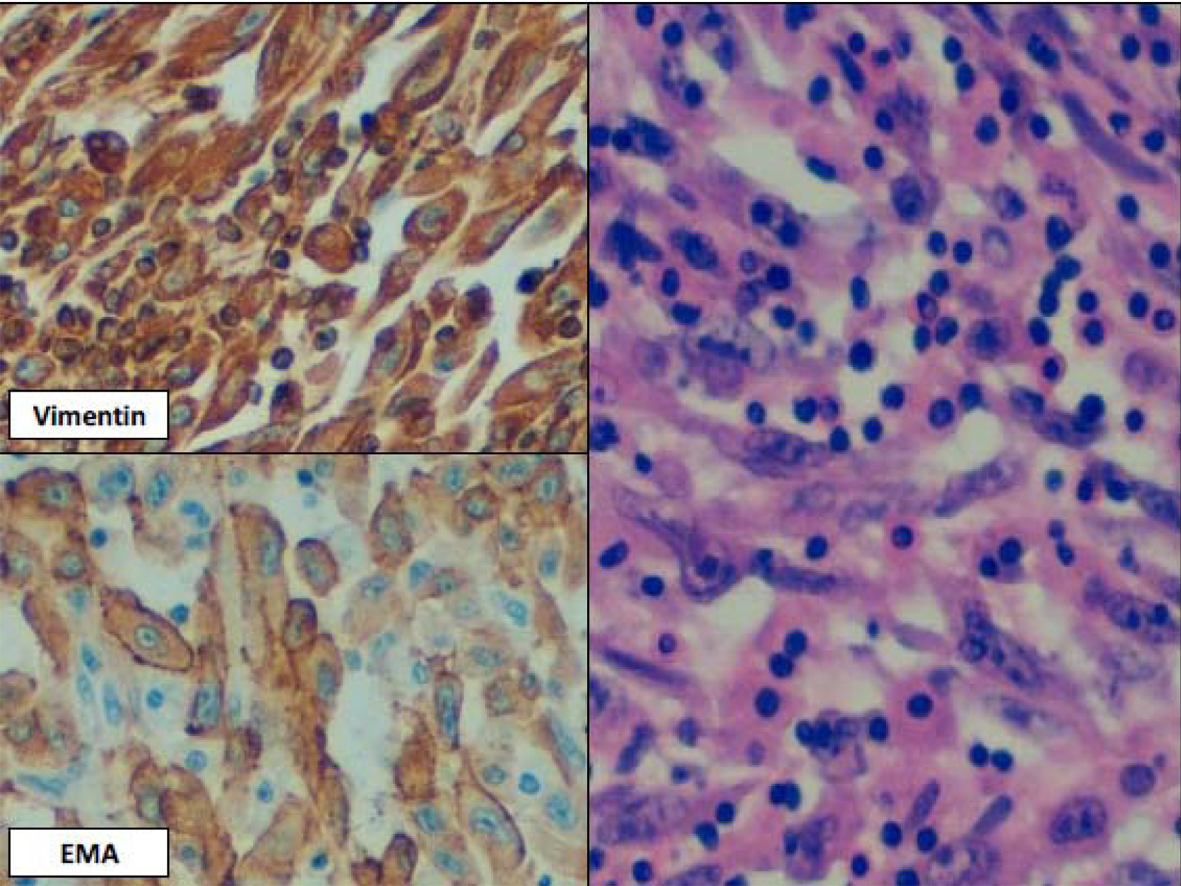
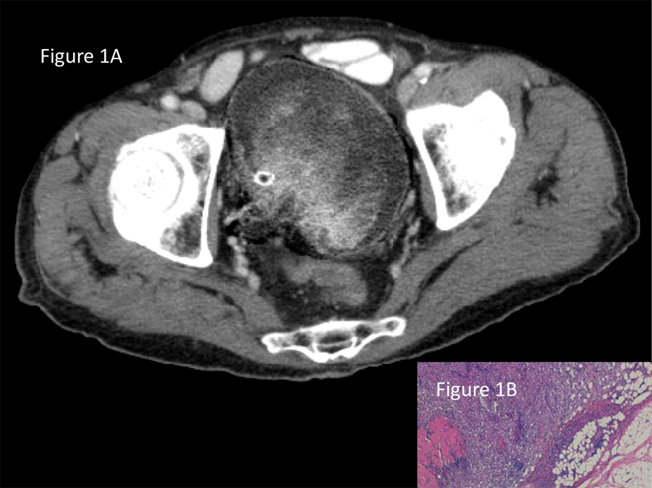
Figure 1. A). CT revealed a large mass occupying almost the entire bladder at an advanced stage disease (T2 or higher) and no metastatic lesions. B). Sarcomatoid carcinoma mostly composed of undifferentiated malignant spindle cells. Tumor infiltrates through adventitial adipose tissue.
Figure 2.
Figure 2. Sarcomatoid carcinoma composed entirely of sarcomatous appearing cells.
Figure 3.
Figure 3. Higher magnification of Fig. 2 with vimentin and EMA positivity documenting epithelial differentiation.

Table

Table 1. Differential Diagnosis of Urinary Bladder Sarcomatoid Carcinoma According to Morphological and Immunohistochemical Features
 
Sarcomatoid CarcinomaLeiomyosarcomaHigh grade urothelial carcinoma with metaplastic changesInflammatory myofibroblastic tumorPost-operative spindle cell nodule
EMA: epithelial membrane antigen; MSA: muscle-specific actin;* Rare aberrant expression. ** Regarding the specific mesenchymal differentiation.
Cytologic atipia in spindle cellsYes (High grade)YesNoNoNo
Concurrent neoplastic epithelial elementsYesNoYesNoNo
AE1/AE3+*+*-
CK 5/6++
P63++
EMA+-+--
Vimentin+++++
MSA*+**-+
Desmin-+**-+