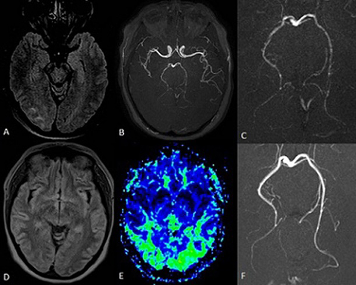
Figure 1. Brain MRI (FLAIR sequences) performed 6 h after symptoms onset showed edema along the occipital subcortical white matter, more evident on the right side (A). MR angiography (B, C) showed a less intense representation of the posterior cerebral arteries with multiple irregularities in their walls (segmental stenoses). The resolution of the edema is seen on the MRI performed 72 h later (D), with signs of reactive hyperperfusion of the posterior regions on perfusion maps (E). An angio-MRI performed 4 weeks later showed the resolution of the posterior circulation segmental stenoses (F).