


| Journal of Medical Cases, ISSN 1923-4155 print, 1923-4163 online, Open Access |
| Article copyright, the authors; Journal compilation copyright, J Med Cases and Elmer Press Inc |
| Journal website http://www.journalmc.org |
Case Report
Volume 4, Number 11, November 2013, pages 769-770
Concussion Induced Electrocardiogram Changes
Michael T Flannery
Department of Internal Medicine, University of South Florid Morsani College of Medicine, 12901 Bruce B. Downs Blvd., MDC Box 19, Room L1041, Tampa, FL 33612, USA
Manuscript accepted for publication June 6, 2013
Short title: Concussion Induced Electrocardiogram Changes
doi: https://doi.org/10.4021/jmc1338w
| Abstract | ▴Top |
A middle aged male presented to the Emergency room with pre-syncope following a hockey induced concussion. Abnormal electrocardiogram changes combined with a strong family history resulted in a cardiac catheterization which was normal. Nearly 2 years later, flattening of the T waves persisted with symptoms. Three years later, the patient developed ALS with predominant upper motor neuron findings.
Keywords: Concussion; Closed brain injury; Abnormal electrocardiogram; Lou Gehrig’s disease (ALS)
| Introduction | ▴Top |
Concussion injuries in sports have received increased attention in prevention not only due to acute and sub-acute symptoms but potentially long-term effects of brain injury including typical features of closed brain injury especially when recurrent. These may include loss of cognitive function effecting activities of daily living similar to dementia. More recently, there are concerns of amyotrophic lateral sclerosis (ALS) or Lou Gehrig’s disease due to recurrent concussions. There are many findings in concussions and our case reviews some of the electrocardiogram changes seen with acute injury. These changes are typically not followed over time and patients may not remember to relate new symptoms to their prior injury.
| Case Report | ▴Top |
A 46-year-old male presented with pre-syncope stating he had struck his head against the ice rink board 2 days previously with some residual headache. His history included a prior concussion at age 12 and a reported normal electrocardiogram (ECG) in 1986 (baseline). The patient was, otherwise, asymptomatic. A complete exam, laboratory studies with cardiac isoenzmes and a computed tomography of the head were normal. An ECG was performed as shown in Figure 1 which demonstrated inverted T waves in V1-V5 and flattening of the T waves in V6, III and aVf and an incomplete right bundle branch block (QRS, 110 ms). Given a strong family history, the patient underwent cardiac catheterization which was normal. The patient reported post-concussion symptoms for 3 months, after the incident, including headache and pre-syncope. A subsequent ECG was performed at 28 months later (Fig. 2), which showed mild flattening of the T waves (QRS, 100 ms).
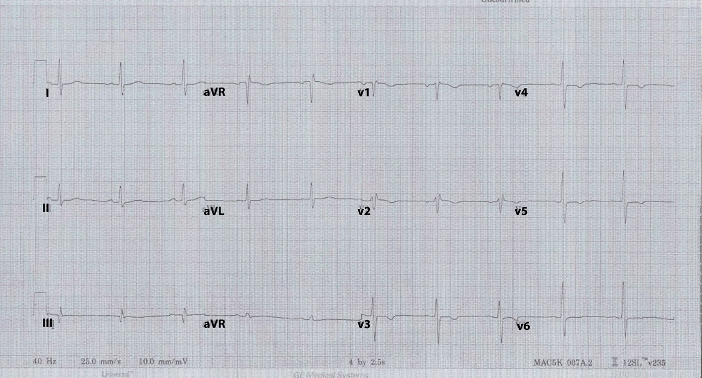 Click for large image | Figure 1. ECG at time of incident demonstrating fairly diffuse T wave inversions. |
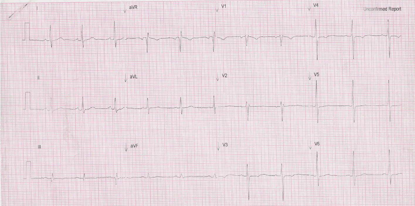 Click for large image | Figure 2. Routine follow-up ECG 28 months later which still shows some T wave changes that have not resolved to a normal state. |
| Discussion | ▴Top |
Concussions causing ECG abnormalities are rarely discussed but have been reported with similar findings [1, 2]. Finally, our patient was diagnosed, 4 years later, with amyotrophic lateral sclerosis which controversially may be associated with head trauma [3-5]. The patient resented with predominately upper motor signs and symptoms such as muscle spasm and hyperreflexia which offer better prognostic findings in the setting of ALS. Given the fact that such long-term side effects can be devastating and life threatening, attention should be focused on prevention and then appropriate diagnosis and treatment which typically involve a lengthy period of inactivity from sports which is obviously frustrating to the sports player. Patients should be honest and forthcoming regarding concussion like symptoms that may persist such as headaches, pre-syncope, ringing in the ears and a sense of imbalance. Without special attention to subsequent prevention, patients may develop more serious long-term effects as previously described. The electrocardiographic changes, as seen in this case, should serve as a reminder to the caring physician, so that unnecessary invasive procedures will not be performed potentially putting the patient at additional serious risks including hematoma, bleeding and infection with the occasional death due to rhythm disturbances or cardiac wall penetration causing tamponade physiology.
| References | ▴Top |
- Ommaya AK. Trauma to the nervous system. Ann R CollSurg Engl. 1966;39(6):317-347.
pubmed - Greenspahn BR, Barzilai B, Denes P. Electrocardiographic changes in concussion. Chest. 1978;74(4):468-469.
doi pubmed - DeKosky ST, Ikonomovic MD, Gandy S. Traumatic brain injury—football, warfare, and long-term effects. N Engl J Med. 2010;363(14):1293-1296.
doi pubmed - Bedlack RS, Genge A, Amato AA, Shaibani A, Jackson CE, Kissel JT, Wall C, et al. Correspondence regarding: TDP-43 proteinopathy and motor neuron disease in chronic traumatic encephalopathy. J NeuropatholExpNeurol 2010:69;918-29. J NeuropatholExp Neurol. 2011;70(1):96-97; author reply 98-100.
doi pubmed - Bartholet J. The collision syndrome.Sci Am. 2012;306(2):66-71.
doi pubmed
This is an open-access article distributed under the terms of the Creative Commons Attribution License, which permits unrestricted use, distribution, and reproduction in any medium, provided the original work is properly cited.
Journal of Medical Cases is published by Elmer Press Inc.